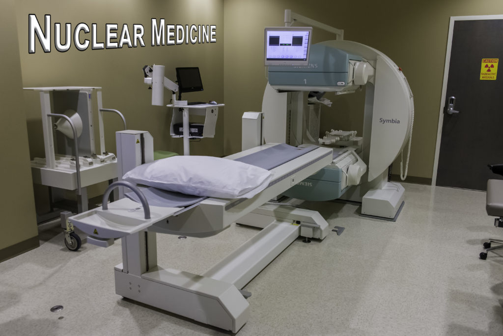
Nuclear medicine is a safe and painless imaging technique used often to help physicians diagnose illness. It is a special branch of radiology that provides a picture of the body’s systems in action.
Are the key organ systems working the way they should? Is brain activity normal? Is the digestive system working properly? Is chronic pain the result of a tiny bone fracture too small to detect?
Nuclear Medicine answers these questions and more with the help of safe, short lived radioactive materials that move through the body and show how the organ systems are functioning.
Nuclear scans or scintigraphy are especially helpful in finding and in some cases treating blockage, locating pockets of infection, identifying hidden fractures, visualizing tumors, and demonstrating changes in bones related to aging or overuse. The scans often locate disease very early in its process when there may be more success in treating it.
Nuclear Medicine
A nuclear medicine procedure is sometimes described as an “inside-out” x-ray because it records radiation emitting from the patient’s body rather than radiation that is directed through the patient’s body. Nuclear medicine procedures use small amounts of radioactive materials, called radiopharmaceuticals, to create images of anatomy.
Radiopharmaceuticals are substances that are attracted to specific organs, bones or tissues. They are introduced into the patient’s body by injection, swallowing or inhalation. As the radiopharmaceutical travels through the body, it produces radioactive emissions.
A special type of camera detects these emissions in the organ, bone or tissue being imaged and then records the information on a computer screen or on film. Nuclear medicine is unique because it documents function as well as structure. For example, nuclear medicine allows physicians to see how a kidney is functioning, not just what it looks like.
Most other diagnostic imaging tests, in comparison, reveal only structure. Nuclear medicine procedures are performed to assess the function of nearly every organ. Common nuclear medicine procedures include thyroid studies, brain scans, bone scans, lung scans, and liver and gallbladder procedures.
Although nuclear medicine is primarily used for diagnosis, it can be used to treat disease as well. Therapeutic uses include treatment of hyperthyroidism and pain relief from certain types of bone cancers.
Before your examination, a nuclear medicine technologist will explain the procedure to you and answer any questions you might have. A nuclear medicine technologist is a skilled medical professional who has received specialized education in the areas of anatomy, radiation protection, patient care, radiation exposure, radiopharmaceuticals and nuclear medicine procedures.
Tell the technologist if you have any allergies and if you are undergoing radiation therapy, because these factors may require adjustments in how the examination is performed. Also, be sure to tell the technologist if you are pregnant or are breastfeeding. Nuclear medicine tests usually are not recommended for pregnant women.
Peachtree City office at (770) 305-4674
Atlanta office at (404) 225-5674
