Bone density scan also frequently called a DEXA or bone mineral density exam, is an important procedure in the evaluation of bone health. The technology uses a dedicated machine  designed specifically for this purpose. It is a quick, simple and painless exam that uses x-rays and highly sophisticated software to measure bone loss.
BMD is our best defense in the early diagnosis and treatment of osteoporosis. Often called the silent disease, osteoporosis robs the body of bone strength, leaving many at risk for broken bones with even a simple fall or bump.
Postmenopausal women are at highest risk but both men and women lose bone strength as they age. In recent years, the emergence of many new pharmaceutical options has greatly enhanced the treatment of osteoporosis. With these new treatment options available, diagnosing bone loss early can have a significant impact on quality of life for osteoporosis patients.
The Advanced Body Composition assessment feature produces color images displaying the distribution of fat, lean mass, bone and fat mass index. The information is translated into an easy-to-interpret report for improved patient management and counseling.
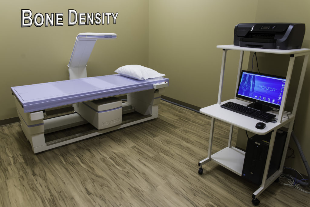
Bone Density Scan
This examination is usually done on an outpatient basis. In the Central DXA examination, which measures bone density in the hip and spine, the patient lies on a padded table. An x-ray generator is located below the patient and an imaging device, or detector, is positioned above.
To assess the spine, the patient’s legs are supported on a padded box to flatten the pelvis and lower (lumbar) spine. To assess the hip, the patient’s foot is placed in a brace that rotates the hip inward. In both cases, the detector is slowly passed over the area, generating images on a computer monitor.
You must hold very still and may be asked to keep from breathing for a few seconds while the x-ray picture is taken to reduce the possibility of a blurred image. The technologist will walk behind a wall or into the next room to activate the x-ray machine. The peripheral tests are simpler.
The finger, hand, forearm or foot is placed in a small device that obtains a bone density reading within a few minutes. An additional procedure called Lateral Vertebral Assessment (LVA) is now being done at many centers. LVA is a low-dose x-ray examination of the spine to screen for vertebral fractures that is performed on the DXA machine.
The LVA test adds only a few minutes to the DXA procedure. The DXA bone density test is usually completed within 10 to 30 minutes, depending on the equipment used and the parts of the body being examined. You will probably be asked to fill out a questionnaire that will help the doctor determine if you have medical conditions or take certain medications that either increase or decrease your risk of a fracture.
The World Health Organization has recently released an online survey that combines the DXA results and a few basic questions and can be used to predict 10-year hip fracture risk for post-menopausal women. This will be coming into more use in the next few years.
Bone density tests are a quick and painless procedure. Expect to be in and out quickly! This painless test only takes about 10 minutes. You will lie flat on your back on a padded table. The DXA (Dual Energy X-ray Absorptiometry) machine scans the spine and hip.
The radiology technician may place a special pillow under your knees for part of the test. Don’t worry about radiation exposure? it is minimal. Routine evaluations every two years may be needed to see a significant change in bone mineral density, decrease or increase. Few patients, such as patients on high dose steroid medication, may need follow-up at six months.
Peachtree City office at (770) 305-4674
Newnan office at (770) 502-9883
Atlanta office at (404) 225-5674