
Ultrasound is a safe, painless diagnostic procedure that uses high-frequency sound waves to see specific areas within the body. It uses the same echo-locating principles of sonar technology employed for decades by ships at sea.
Sound waves directed into the body produce echoing waves as they bounce against the internal fluids of the human body. The echoes are captured and reconstructed by a sophisticated computer software into live images on the computer monitor.
The primary benefit of ultrasound is its ability to generate highly-detailed images without being invasive and without using radiation. Because it is considered so safe and simple, ultrasound has become the diagnostic tool of choice to examine the well-being of unborn children as well as many other specific exams.
Ultrasound
OPI offers a full array of ultrasound examinations to evaluate and diagnose abdominal, gynecological, obstetrical, fetal, pediatric, thyroid, breast, scrotal, prostate and musculoskeletal conditions, as well as vascular pathology.
For pregnancy, ultrasound can help determine gestational age and evaluate anatomical development. It is also used to screen for the risk of Down syndrome and other genetic abnormalities in the first trimester.
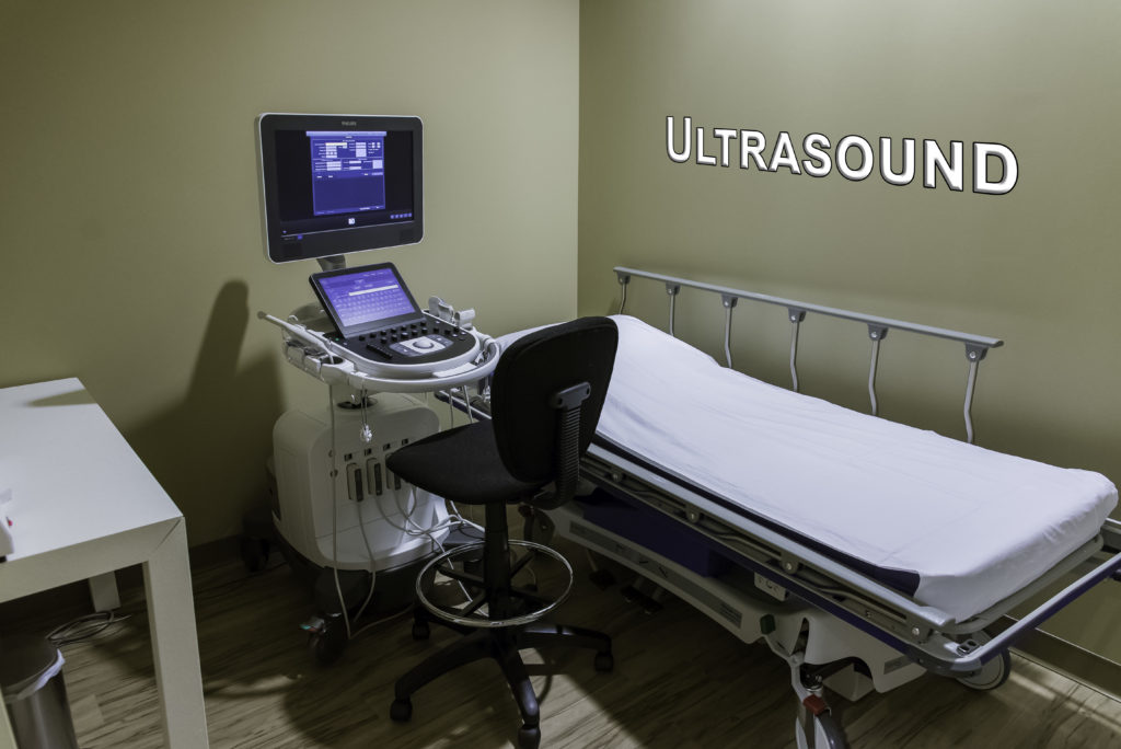
Equipment for an ultrasound consists of a console containing a computer and electronics, a video display screen and a transducer, which is a small, hand-held device used to scan the body. The transducer distributes high frequency sound waves into the body and then listens for the return echo from the tissues in the body. This return echo is used to form the ultrasound image, which is immediately visible on the computer screen.
Patients lie on an examining table that may be tilted with the head up. A clear gel is applied to the area of the body being studied to enable sound waves to travel between the transducer and the patient. The transducer is held firmly by the sonographer or radiologist and moved over the area of interest. Once the examination is complete, patients may be asked to dress and wait while the ultrasound images are reviewed. Most ultrasound examinations are conducted within 30 to 45 minutes.
At OutPatient Imaging, patients are seen by dedicated, fellowship-trained radiologists, who are some of the finest in the field. They maintain a close rapport with referring physicians and are sensitive to the patients’ concerns, taking care to explain each procedure that will be performed. Plus our technical staff of registered diagnostic medical sonographers ensures that patients are at ease during examinations.
Peachtree City office at (770) 305-4674
Newnan office at (770) 502-9883
Atlanta office at (404) 225-5674
![]()